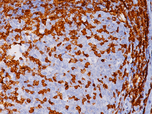
Description
CD3 is expressed throughout the T-cell differentiation process. CD3 is a highly specific and sensitive T-cell lineage marker, making it ideal for the immunophenotypic analysis of lymphohaematopoietic malignancies. Notable exceptions include some of the more aggressive large T-cell lymphomas and CD30 (Ki-1) positive anaplastic large cell lymphomas, which may not express detectable antigen (1-3). CD3 [LN10] has demonstrated optimal staining when compared to other CD3 clones including PS1, F7.2.38 and SP7 (4). A monoclonal antibody to human CD3 is regarded as a reliable pan T-cell antibody used in the immunophenotyping of lymphomas in paraffin sections.
SPECIFICATIONS
Specifications
| INTENDED USE | IVD |
|---|---|
| FORMAT | Concentrate, Predilute, VALENT |
| VOLUME | 0.1 ml, 1.0 ml, 20 ml, 25 ml, 6.0 ml |
| SOURCE | Mouse Monoclonal |
| CLONE | LN10 |
| ISOTYPE | IgG1 |
| ANTIGEN | CD3 |
| LOCALIZATION | Cell surface |
| POSITIVE CONTROL | Tonsil |
DATASHEETS & SDS
REFERENCES
1. Campana D, et al. The cytoplasmic expression of CD3 antigens in normal and malignant cells of the T lymphoid lineage. J Immunol. 1987 Jan; 138(2):648-55.
2. Cabeçadas JM, Isaacson PG. Phenotyping of T-cell lymphomas in paraffin sectionswhich antibodies? Histopathology. 1991 Nov; 19(5):419-24.
3. Steward M, et al. Production and characterization of a new monoclonal antibody effective in recognizing the CD3 T-cell associated antigen in formalin-fixed embedded tissue. Histopathology. 1997 Jan; 30(1):16-22.
4. “CD3 Assessment Run 37 2013.” NordiQC. NordiQC, 04 Dec. 2013. Web. 16 June 2015.
5. Center for Disease Control Manual. Guide: Safety Management, NO. CDC-22, Atlanta, GA. April 30, 1976 “Decontamination of Laboratory Sink Drains to Remove Azide Salts.”
6. Clinical and Laboratory Standards Institute (CLSI). Protection of Laboratory Workers from Occupationally Acquired Infections; Approved Guideline-Fourth Edition CLSI document M29-A4 Wayne, PA 2014.


Reviews
There are no reviews yet.