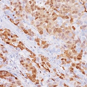
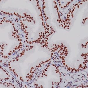
Description
p63 has been shown to be a sensitive marker for lung squamous cell carcinomas (SqCC), with reported sensitivities of 80-100% (1-6). Specificity for lung SqCC, versus lung adenocarcinoma (LADC), has been reported to be approximately 70-90%, as positive staining with p63 has been typically observed in 10-30% of LADC (1-6). Cocktails of p63 with complementary markers for lung SqCC have also proven useful. A cocktail of p63 TRIM29 demonstrated a 94.7% sensitivity for lung SqCC and 100% specificity versus LADC, in cases where Napsin A and TTF-1 were both negative (2-7). Similarly, the combination of p63 CK5 identified 87% of cases of lung SqCC, with 94% specificity (8).
SPECIFICATIONS
Specifications
| INTENDED USE | IVD |
|---|---|
| FORMAT | Predilute |
| VOLUME | 6.0 ml |
| SOURCE | Mouse Monoclonal |
| CLONE | 4A4 |
| ISOTYPE | IgG2a /kappa |
| ANTIGEN | p63 |
| LOCALIZATION | Nuclear |
| POSITIVE CONTROL | Lung squamous cell carcinoma |
| SPECIES REACTIVITY | Human, Mouse, Rat |
REFERENCES
1. Mukhopadhyay S, Katzenstein AL. Subclassification of non-small cell lung carcinomas lacking morphologic differentiation on biopsy specimens: Utility of an immunohistochemical panel containing TTF-1, napsin A, p63, and CK5/6. Am J Surg Pathol. 2011 Jan; 35(1):15-25. 2. Tacha D, et al. A Six Antibody Panel for the Classification of Lung Adenocarcinoma versus Squamous Cell Carcinoma. Appl Immunohistochem Mol Morphol. 2012 May; 20(3):201-7. 3. Kargi A, Gurel D, Tuna B. The diagnostic value of TTF-1, CK 5/6, and p63 immunostaining in classification of lung carcinomas. Appl Immunohistochem Mol Morphol. 2007 Dec; 15(4):415-20. 4. Khayyata S, et al. Value of P63 and CK5/6 in distinguishing squamous cell carcinoma from adenocarcinoma in lung fine-needle aspiration specimens. Diagn Cytopathol. 2009 Mar; 37:178–83. 5. Terry J, et al. Optimal immunohistochemical markers for distinguishing lung adenocarcinomas from squamous cell carcinomas in small tumor samples. Am J Surg Pathol. 2010 Dec; 34(12):1805-11. 6. Pu RT, Pang Y, Michael CW. Utility of WT-1, p63, MOC31, mesothelin, and cytokeratin (K903 and CK5/6) immunostains in differentiating adenocarcinoma, squamous cell carcinoma, and malignant mesothelioma in effusions. Diagn Cytopathol. 2008 Jan; 36(1):20-5. 7. Tacha D, Yu C, Haas T. TTF-1, Napsin A, p63, TRIM29, Desmoglein-3 and CK5: An Evaluation of Sensitivity and Specificity and Correlation of Tumor Grade for Lung Squamous Cell Carcinoma vs. Lung Adenocarcinoma. Mod Pathol. 2011 Feb; 24 (Supplement 1s):425A. 8. Tacha D, Zhou D, Henshall-Powell RL. Distinguishing Adenocarcinoma from Squamous Cell Carcinoma in Lung Using Double Stains p63+ CK5 and TTF-1 + Napsin A. Mod Pathol. 2010 Feb; 23 (Supplement 1s):222A. 9. Center for Disease Control Manual. Guide: Safety Management, NO. CDC-22, Atlanta, GA. April 30, 1976 “Decontamination of Laboratory Sink Drains to Remove Azide Salts.” 10. Clinical and Laboratory Standards Institute (CLSI). Protection of Laboratory workers from occupationally Acquired Infections; Approved guideline-Third Edition CLSI document M29-A3 Wayne, PA 2005.


Reviews
There are no reviews yet.